
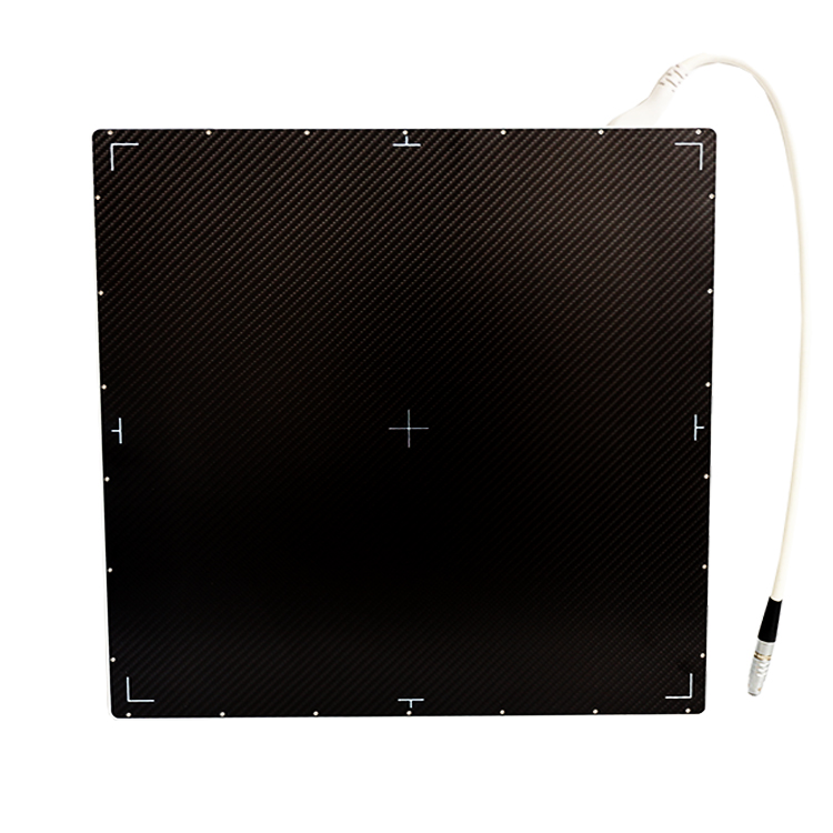
Flat panel detectors have revolutionized the field of medical imaging by providing high-quality images with minimal radiation exposure. Among the various flat panel detector technologies, amorphous selenium flat panel detectors stand out due to their unique working principle and superior image quality.
Amorphous selenium flat panel detectors utilize a thin layer of amorphous selenium as the photoconductive material. When X-rays pass through the patient and reach the detector, they are absorbed by the selenium layer, creating electron-hole pairs. These charged carriers are then drifted towards the electrodes located at the top and bottom of the detector, creating an electric signal that is proportional to the X-ray intensity.
One of the key advantages of amorphous selenium detectors is their direct conversion of X-rays into electrical signals. This direct conversion process eliminates the need for scintillators or other intermediate materials, resulting in higher spatial resolution and improved image quality. Additionally, amorphous selenium’s high atomic number and density make it an efficient absorber of X-rays, further enhancing the detector’s sensitivity.
In the absence of an electric field, the electron-hole pairs in amorphous selenium tend to recombine, leading to signal decay and loss of image quality. To prevent this, amorphous selenium detectors are equipped with a bias voltage that creates an electric field, separating the charged carriers and allowing them to reach the electrodes without recombining.
The bias voltage, typically in the range of 5-10 kV, is applied to the electrodes during image acquisition, ensuring that the electric field is constantly present to maintain signal integrity. This continuous charge collection process facilitates fast image acquisition, making amorphous selenium detectors suitable for real-time imaging applications such as fluoroscopy and interventional procedures.
Moreover, amorphous selenium’s stable and robust nature allows for long-term reliability and minimal maintenance requirements, making it an ideal choice for medical imaging systems. The direct conversion and signal amplification capabilities of amorphous selenium detectors result in low noise and high detective quantum efficiency (DQE), contributing to excellent image contrast and visibility of anatomical details.
In addition to medical imaging, amorphous selenium flat panel detectors have found applications in industrial non-destructive testing and security screening due to their high-performance characteristics. Their ability to produce high-resolution, low-noise images in real-time makes them invaluable tools in a wide range of imaging scenarios.
As technology continues to advance, the potential for further improvements in amorphous selenium flat panel detectors is vast. Ongoing research aims to enhance their performance by optimizing the charge transport mechanisms, refining the design of the electrodes, and exploring new materials for the detector structure.
Overall, the working principle of amorphous selenium flat panel detectors, coupled with their exceptional image quality and reliability, underscores their significance in advancing the field of medical imaging and beyond. As the demand for high-quality, low-dose imaging solutions continues to grow, amorphous selenium detectors are poised to play a pivotal role in shaping the future of radiology and imaging sciences.
Post time: Aug-06-2025