

Against the backdrop of rapid advancement within digital radiography (DR) ecosystems, one pivotal component governs image fidelity, diagnostic throughput and radiation hazard mitigation — the Flat Panel Detector (FPD). Hailed as the beating core of DR apparatus, it underpins the paradigm shift away from analogue X-ray film toward full digitalisation. This discourse dissects this indispensable module underpinning contemporary medical radiography.
1 What Constitutes a Flat Panel Detector
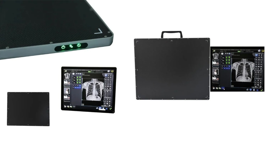
A flat panel detector represents a sophisticated optoelectronic radiographic sensor that captures X-ray projections via a two-dimensional pixel matrix. Simply characterised, it supersedes traditional X-ray cassettes and analogue film, transducing attenuated X-ray fluxes traversing subjects straight into digitised visual signals for rendering on computational terminals. This seminal technological leap surmounts inherent drawbacks of film-based radiography, enabling sub-second image acquisition, minimised radiation exposure and advanced algorithmic post-processing.
2 Operating Mechanisms Two Distinct Paradigms Converting X-Rays to Digital Visuals
Indirect Conversion Architecture exemplified by amorphous silicon
This architecture remains the most ubiquitously deployed commercial solution. X-ray quanta first impinge upon scintillating substrates, typically Caesium Iodide (CsI) or Gadolinium Oxysulfide (GOS), which convert incident radiation into visible luminescence. Subsequently, amorphous silicon photodiode arrays harvest such photons and transform them into analogue electrical impulses; analogue-to-digital conversion (A/D) ultimately generates rasterised digital imagery. Owing to this sequential transduction chain of radiation, luminescence and electric signals, the configuration is termed indirect conversion.
Direct Conversion Architecture exemplified by amorphous selenium
This avant-garde scheme eliminates intermediate luminescence conversion. Incoming X-ray photons directly bombard amorphous selenium (a-Se) strata to spawn electron-hole pairs. Subject to an extrinsic electric field, charge carriers generate electric currents retained within Thin-Film Transistor (TFT) capacitors, yielding digital images post analogue-to-digital transformation. Exclusion of optical transduction circumvents photon scattering, granting superior spatial resolution, ideally suited for high-precision radiography such as mammography and orthopaedic skeletal scanning.
3 Transmission Topologies
Wireless Flat Panel Detector
Unshackled from cabling restraints, wireless variants facilitate unrestricted relocation and angular adjustment, perfectly suited for emergency wards, bedside diagnostics and field deployments. Nevertheless, signal attenuation risks persist, accompanied by elevated commercial pricing.
Wired Flat Panel Detector
Interfaced via dedicated physical cabling, wired detectors deliver robust signal transmission, expedited data throughput and cost competitiveness, albeit constrained mobility radius.
4 Pioneering Medical Provisions Through Refined Engineering Delivering Precise Radiography for Public Wellness
Portable DR equipment transcends the definition of mere mobile imaging hardware. It functions as an instrumental catalyst for grassroots healthcare modernisation, public health outreach and contingency clinical interventions. It emancipates high-grade radiographic diagnosis from fixed radiological suites, extending specialist care beyond hospitals to grassroots institutions and on-site scenarios. Every radiographic examination attains heightened efficiency, accuracy and accessibility, democratising premium diagnostic services.
5 Multifarious Deployment Scenarios
The utilisation scope of flat panel detectors stretches far beyond conventional radiological departments:
- Medical Radiography: DR chest screening, orthopaedic diagnostics, mammography, digital angiography and Cone Beam CT (CBCT).
- Bedside & Emergency Intervention: Urgent radiological assessment for critically ill ICU inpatients and trauma casualties.
- Industrial Non-Destructive Assay: Defect detection within metallic castings and welded joints.
- Security Inspection: Luggage tomography and freight screening protocols.
6 Epilogue
As the foundational hardware enabling digital X-ray radiography, flat panel detectors are catalysing sweeping transformations spanning clinical diagnostics, industrial assay and security surveillance. The industry witnesses continuous evolution: from analogue film to digital sensor panels, from single static exposure to real-time dynamic radiography, and from wired linkage to untethered mobility. Each iterative upgrade renders X-ray imaging sharper, safer and more expeditious. Comprehension of detector operational physics and architectural classifications empowers rational decision-making throughout equipment procurement and clinical manipulation, fostering precise diagnostics and elevated medical standards.
Post time: Jul-22-2026