

Should a small orthopedic clinic invest in a handheld X‑ray unit dedicated to extremity imaging? A handheld X‑ray device built for extremities perfectly fills a gap in imaging demands for small clinics. This is a common scenario in orthopedic clinics: a patient arrives with an ankle sprain. The clinician has a strong clinical suspicion, yet a definitive conclusion cannot be reached without radiographs. Patients are referred to larger hospitals several kilometers away for imaging. After the hassle of round trips, many patients will simply drop out.
It is not that the clinician lacks diagnostic skills, but that there is no radiographic evidence to support assessment. Diagnostic confidence, patient trust, follow‑up visits and referrals all hinge on this missing piece. This article clarifies why a handheld extremity X‑ray unit addresses this specific pain point for small orthopedic practices. It covers what this equipment can deliver, its limitations, and how to decide whether it is a suitable purchase.
01 The core challenge is not the need for imaging, but unaffordable large‑scale systems
The value of radiographic imaging for orthopedics is widely recognized. The real barrier for small clinics lies in the high cost of establishing imaging capacity. Conventional full‑size DR systems carry high price tags, require substantial floor space, dedicated shielded rooms and renovation work, often costing tens of thousands. Most small clinics rule out this option after budgeting — not because imaging is unnecessary, but because the investment is burdensome and the machine may end up underutilized.
Still, skipping in‑house imaging creates hidden drawbacks: clinical assessments rely only on physical palpation; patients feel uncertain without in‑clinic images; follow‑up management and patient referrals lack intuitive visual proof. The question is never whether imaging matters. It is about finding an affordable way to access it.
02 What a handheld extremity X‑ray unit is, and what it is not
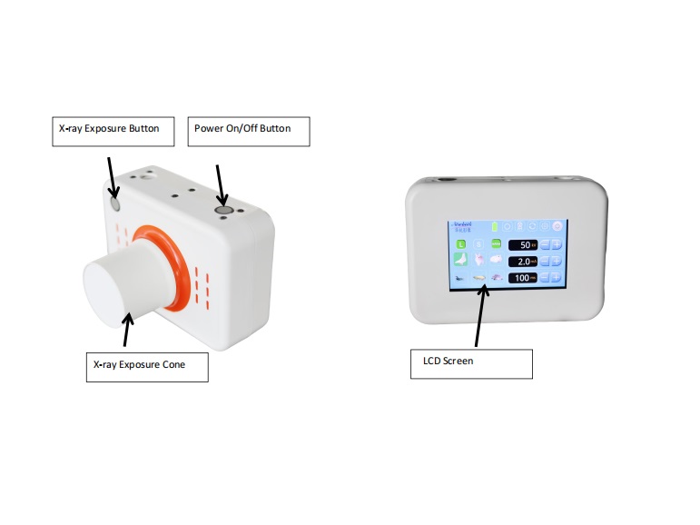
Simply put, this is a portable digital X‑ray system that can be carried and operated directly at the point of care. It is designed for extremities including hands, wrists, elbows, feet, ankles and knees. It is not a replacement for full‑function DR units. It is a specialized tool focused on extremity trauma. It cannot capture full‑body scans or serve all general radiography needs. It also cannot substitute professional clinical judgment; radiographs still require interpretation by qualified practitioners.
Its key transformation is straightforward: extremity radiography no longer requires trips to external radiology suites or large hospitals. Imaging can be completed within the consultation room.
03 It cuts three major categories of operational cost
When evaluating equipment, value is measured not by feature count, but by workflow improvements. Space cost: No dedicated shielded radiographic room is mandatory. Unlike fixed DR equipment that demands specially modified enclosures, handheld systems have flexible site requirements and fit well inside compact clinics. Financial barrier: The entry cost is far lower. Compared with large radiography systems, handheld units demand a much lighter capital outlay. For small clinics, starting with a fit‑for‑purpose portable device is a far more practical strategy than committing to a high‑end complete setup at once. Workflow cost: Imaging is integrated directly into consultations. Patients do not need to travel elsewhere for scans. Images are available instantly, forming a closed loop for consultation and assessment.
04 It covers most high‑frequency orthopedic examinations
Most routine orthopedic caseloads center on extremity conditions: Hands and wrists: Fall injuries, workplace trauma and distal radius fractures, one of the most common fracture types. Elbows: Paediatric dislocations and post‑trauma joint evaluation for adult patients. Ankles: High‑incidence sprain cases to rule out fractures and associated soft tissue injury. Knees: Degenerative changes and preliminary assessment after trauma. Post‑operative follow‑up: Monitoring fracture healing and verifying implant positioning.
All these cases share similar traits: localized extremity pathology, potentially limited patient mobility, and a requirement for immediate preliminary evaluation. These are exactly the scenarios where handheld X‑ray units excel.
05 Four criteria to decide whether to purchase
Instead of judging if handheld X‑ray machines are universally good, assess your clinic’s actual gaps. Check four practical conditions: Do you receive a steady stream of extremity and trauma cases, with frequent wrist, hand, elbow and ankle consultations? Do you often lose patients or delay care because they have to seek imaging off‑site? Can your space and budget accommodate a large fixed DR system? If not, a handheld unit may be a practical first step.
Equipment selection is not about chasing the most expensive or fully featured model. The best device reliably handles your highest‑volume clinical demands.
Post time: Oct-28-2026