

Taking your parents to the hospital, the doctor says “we need an X-ray first”. You agree out loud, but worry deep down: radiation! Is it harmful to the body? Can elderly people tolerate it? Is it safe for children?
This concern is completely reasonable. After all, the terms “X-ray” and “radiation” sound intimidating.
But here is a fact many people overlook: every person is surrounded by natural radiation every single day. From cosmic rays to soil, air and food, an average person receives roughly 2 to 3 millisieverts of natural background radiation each year. This baseline exposure exists for everyone living on Earth; it is unavoidable and nothing to panic about.
So how much radiation comes from one X-ray shot? A chest radiograph delivers approximately 0.02–0.1 millisieverts. To put it simply: the radiation from one chest X-ray is comparable to the exposure from a multi-hour flight, or around ten days of ordinary daily life.
If the dose is this low, why do so many people still feel anxious? Humans naturally fear things invisible and intangible. Misleading online claims also fuel worry: “X-rays cause cancer”, “you cannot get two scans within six months”, “X-ray stunts children’s growth”. These rumours spread widely and create unnecessary alarm.
The truth is that radiation doses for medical X-ray examinations are strictly capped by international safety standards. The exposure from an occasional scan is far below the threshold that poses meaningful risk. The key risk to watch for is frequent, unnecessary repeated imaging — and standardized clinical rules are designed specifically to prevent this.
In short: get the scan when clinically indicated. Clinicians will not order X-rays without valid reasons. The risk is not from one single scan, but from excessive unneeded examinations.
Many people do not realize that the biggest advancement in radiography over the past decade is not sharper image quality. It is the ability to produce clear diagnostic images using far lower radiation doses.
Two major developments make this possible. First, digital imaging has replaced film-based radiography. Old film systems required higher radiation to avoid grainy, faint images. Modern digital detectors are highly sensitive, so they can form usable images with minimal exposure, and eliminate chemical film processing entirely.
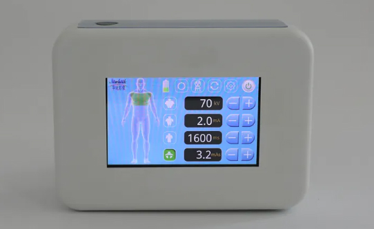
Second, imaging equipment has become compact and mobile. In the past, wrist imaging required a trip to a dedicated radiology suite. Today lightweight handheld X-ray units are available. These portable devices weigh only a few kilograms, enabling extremity imaging directly inside consulting rooms. They are engineered for ultra-low dose and deliver instant image results.
For ordinary people, this means radiography is becoming far less stressful and troublesome. No long trips to specialized departments, no waiting for film development, and lower radiation burden.
Instead of fearing radiation, keep three key points in mind. First, clinicians order imaging after careful risk-benefit assessment. Speak with your doctor about concerns rather than worrying without evidence. Second, the dose of a single diagnostic scan is similar to exposure on a long-distance flight and does not require excessive anxiety. Third, avoid repeated scans without valid medical indication. Qualified practitioners prioritize this just as much as patients.
Understanding radiation helps you make sensible choices for your health, rather than giving in to unfounded fear.
Post time: Nov-20-2026