

For radiographers, clinic operators and medical equipment distributors, mastering the accurate placement of X-ray anti-scatter grid is the key to acquiring crisp, artifact-free radiographic images. Improper grid layout will lead to blurred films, uneven brightness, reduced diagnostic accuracy, and even irreversible damage to imaging devices. This article interprets the placement standards of X-ray grid from multiple dimensions, including working principle, core placement rules, applicable scenarios and on-site operation taboos, helping medical practitioners standardize daily radiography work.
1. Basic Working Principle of X-ray Grid
Before learning placement skills, it is necessary to clarify the core function of the anti-scatter grid. When X-rays penetrate human or animal tissues, primary useful rays pass straight through and project onto flat panel detectors or X-ray films to form valid anatomical images. Meanwhile, scattered secondary X-rays will be generated inside the human body; these disordered scattered rays have no diagnostic value and will overlay on the image, causing overall gray fog, lowering contrast and obscuring tiny lesions such as subtle fractures and pulmonary nodules.
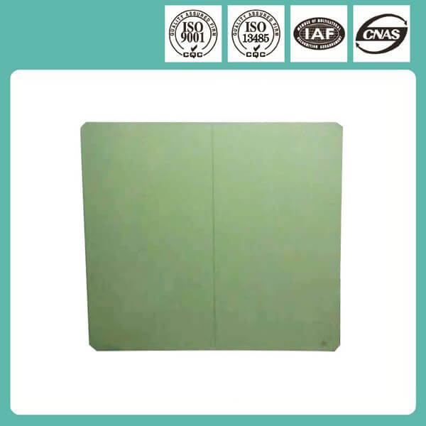
An X-ray grid consists of countless parallel lead strips and interspersed aluminum or fiber spacers. Straight useful X-rays can smoothly pass through the gaps between lead strips, while disorderly scattered rays will be blocked and absorbed by lead strips. The grid converts messy scattered radiation into clear imaging contrast, which is the fundamental basis for its placement requirements.
2. Core Placement Standard: The Focal Side Must Face the X-ray Tube
This is the most critical rule for grid installation, and also the most frequent error in clinical operation. Each X-ray grid has two distinct sides: the focal side and the image receiving side. The focal side, marked with a focal line or “FOCUS” logo, must be placed toward the X-ray tube and X-ray source; the flat reverse side faces the flat panel detector or imaging plate.
The lead strips inside the focused grid are designed to incline slightly toward the X-ray focal spot. Only when the focal side faces the tube can the gap angle of lead strips match the emission angle of primary X-rays. If placed reversely, primary rays will be intercepted by lead strips in large quantities, resulting in extremely dark, unusable images with obvious black strip shadows.
3. Distance Matching: Strictly Conform to Rated Focal Distance Range
Every filter grid is labeled with a rated focal distance, such as 100cm, 150cm, 180cm, which refers to the valid distance between the X-ray tube focal spot and the grid surface. If the actual shooting distance is shorter or longer than the calibrated range, the lead strip angle cannot match the ray angle, causing marginal vignetting: the middle part of the image is clear, while both sides gradually darken. For portable handheld DR and bedside radiography, users should select short focal distance grids; fixed chest DR rooms mostly adopt 180cm long focal distance grids for upright chest examinations.
4. Horizontal & Vertical Placement: Distinguish Linear Grid and Cross Grid
Linear grids are the most widely used type in clinics, only blocking scattered rays along one direction. During limb, chest and abdominal radiography, linear grids need to be placed with lead strips parallel to the long axis of the human body, which can maximally filter scattered rays generated from thick body parts. Cross grids feature dual-direction lead strips, applicable for thick torso scanning; they have no strict horizontal or vertical limits but demand stricter focal distance control, mostly used in large fixed DR systems.
5. Practical Operation Notes & Scenario Selection
Thin body parts such as hands, wrists and calves produce few scattered rays, and no grid is required for shooting; grids are mandatory for torso, abdomen, pelvis and thick limb examinations to guarantee image quality. In veterinary radiography, large dogs and livestock torso shooting also need matched grids for installation.
After placement, operators need to double-check the focal side mark, shooting distance and horizontal direction before exposure. Correct grid placement effectively cuts retake rates, reduces repeated radiation exposure for patients, and prolongs the service life of flat panel detectors.
Conclusion
The proper placement of X-ray grid relies on the combination of principle cognition and standardized operation. Clarifying the focal side direction, rated focal distance matching and directional laying rules can eliminate scattered radiation interference, deliver high-definition diagnostic images, reduce clinical errors, and bring tangible efficiency and safety benefits to human and veterinary medical imaging work.
Post time: Aug-14-2026