

Veterinary radiology has long relied on fixed X-ray rooms inside clinics, which creates hurdles for large livestock, stressed pets, and field emergency cases. The handheld vet X-ray machine changes this workflow, bringing radiographic imaging directly to animals instead of transporting patients to the exam room. Compact, lightweight and portable, this device has quickly become a staple tool for mobile vet services, farm animal checkups and equine field diagnosis.
How Handheld Vet X-Ray Works
Just like stationary X-ray generators, handheld vet X-ray machines use high-frequency X-ray tube technology. When the operator triggers exposure, the generator releases controlled X-ray photons. These photons penetrate animal tissues. Denser structures such as bones absorb more radiation, while soft tissues like organs, skin and muscle allow more rays to pass through. The flat-panel DR detector captures the residual radiation and converts the signal into a digital image instantly.
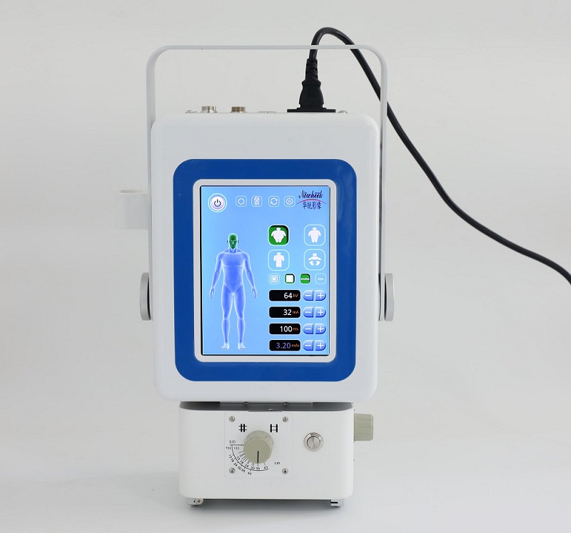
The core difference lies in its miniaturized high-frequency generator. Traditional fixed X-ray units are bulky and need hardwired mains power. Handheld models pack the generator, control panel and X-ray tube into one portable unit. Many versions can run on rechargeable batteries, so vets can work at stables, farms or remote sites without access to wall power.
It is worth noting that handheld vet X-ray does not reduce the importance of radiation safety. Even though the machine is small, X-rays are ionizing radiation. Operators must use proper shielding, maintain safe distances and follow local radiation regulatory rules during every exposure.
Core Features of Handheld Vet X-Ray
Portability is the most prominent feature. Most handheld vet X-ray units weigh only a few kilograms. Veterinarians can carry them in protective carry cases to farm sites, mobile clinics or rescue locations. There is no requirement for dedicated X-ray room construction.
Flexible kVp and mA adjustment is another key advantage. Operators can tune tube voltage and current according to animal size and body thickness. For small animals such as cats, small dogs and rabbits, lower kV settings are used to capture fine soft tissue detail. For bigger animals including sheep, goats and horses, higher kV values penetrate thicker body tissue for clear bone radiographs.
Most modern handheld vet X-ray systems pair with digital flat panel detectors. Images transfer wirelessly to laptops or tablets within seconds. No film processing is needed. Vets can view radiographs right on site, evaluate fractures, joint damage or foreign bodies immediately, and share images with clients or specialist veterinarians quickly.
Practical Value for On-Site Animal Radiology
Moving large or injured animals is risky. Horses, cattle and severely traumatized pets may get stressed, hurt further during transportation. Handheld vet X-ray eliminates this risk by bringing imaging equipment to the animal’s location. This is one of its biggest practical benefits for field veterinary practice.
For mobile vet businesses, this machine also lowers setup investment. Building a dedicated X-ray suite with radiation shielding in a clinic costs heavily. Handheld devices allow new veterinary services to launch radiographic capability with much lower upfront capital. It also adds new service opportunities, such as farm herd screening and pre-purchase equine examinations.
However, users need to understand its limitations. Handheld generators usually have lower power output than fixed clinic X-ray systems. They are best suited for extremity imaging — legs, hooves and joints. For full torso scans of very large animals, fixed high-power X-ray equipment remains a better option. Operators also need training on exposure parameter selection. Wrong kVp or mA settings will produce noisy, low-quality images that are hard to interpret.
Tips to Get Reliable Field Radiographs
Successful on-site imaging depends on a few simple operational habits. First, stabilize the animal as much as possible to reduce motion blur. Even slight movement will blur radiographs and affect diagnosis. Second, keep consistent source-to-image distance. Changing the distance will alter image density and magnification. Third, use a suitable anti-scatter grid when imaging thicker animal parts. The filter cuts scattered radiation, boosts contrast and improves image clarity.
Always plan radiation protection before exposure. Keep people out of the primary beam path, use lead aprons and lead gloves where required. Check local veterinary radiation compliance laws, as many regions require registration and operator certification for portable X-ray devices.
Wrap Up
Handheld vet X-ray fills an important gap in mobile animal radiology. By combining compact high-frequency X-ray generation and fast digital imaging, it lets veterinarians capture diagnostic-quality radiography on location. It works wonderfully for mobile vet clinics, equine practices and livestock farms. When used correctly with proper safety protocols and parameter tuning, it delivers fast diagnostic results and improves animal welfare by avoiding the stress of patient transport. As field veterinary services keep growing, handheld vet X-ray will remain an essential tool for modern on-site animal radiology.
Post time: Sep-28-2026