

Handheld vet X-ray cameras have transformed veterinary radiography, bringing portable, high-definition imaging directly to animal patients. Unlike bulky fixed X-ray systems confined inside examination rooms, this compact device allows vets to perform radiographic exams on-site, which is especially valuable for large, anxious, or critically ill pets. This article explains its working principle, core advantages, practical application tips and key considerations for veterinary clinics.
How Handheld Vet X-Ray Camera Works
At its core, the handheld vet X-ray camera relies on the basic property of X-rays: X-ray photons penetrate animal tissues, and different tissues absorb rays at varying rates. Dense structures like bones block most X-rays, while soft tissues such as skin, muscle and organs allow more rays to pass through.
When the device is triggered, the X-ray tube inside generates controlled X-ray beams. The beam passes through the pet’s body and reaches a digital flat panel detector placed behind the animal. The detector converts the remnant X-ray signal into digital data, and the built-in imaging system instantly processes the data to form a high-definition radiographic image.
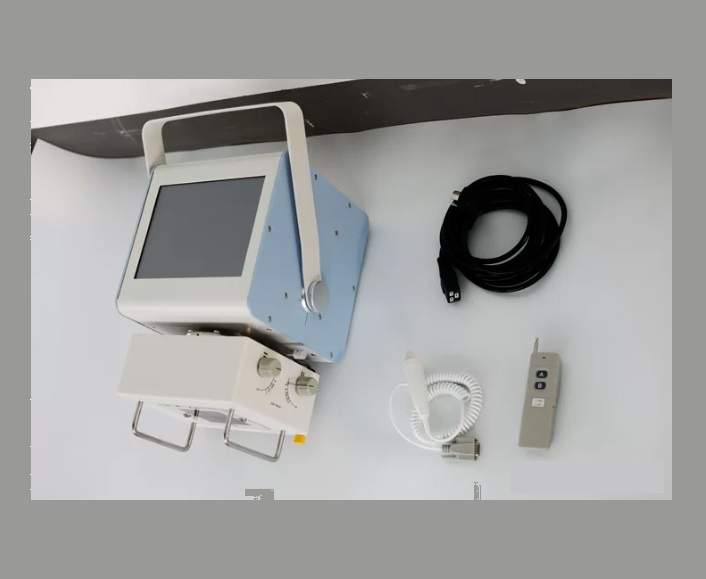
Different from traditional stationary X-ray machines, the handheld unit integrates the X-ray generator, control panel and safety shielding into one lightweight body. Vets can adjust exposure parameters including kV and mAs directly on the device according to the pet’s size and body thickness. This real-time parameter adjustment guarantees clear images for cats, dogs, small mammals and even exotic pets.
What Makes High Definition Imaging Possible for Pets
High-definition image quality is the biggest highlight of modern handheld vet X-ray cameras, and it comes from multiple technical upgrades. First, optimized X-ray tube design delivers stable and consistent beam output, reducing image noise caused by unstable radiation. Second, matched digital flat panel detectors adopt high-sensitivity sensors, which capture subtle differences in tissue density. Tiny lesions, fine bone fractures and soft tissue abnormalities can be displayed clearly on the final image.
Scattered radiation control also plays a vital role in image clarity. Scatter rays create gray fog on images and obscure details. Many handheld vet X-ray cameras support matching portable grid accessories. The filter grid absorbs scattered rays and keeps only useful primary X-rays hitting the detector, greatly improving image contrast.
For small pets, overexposure or underexposure is a common problem. The handheld camera’s automatic exposure compensation function solves this pain point. It automatically fine-tunes radiation output for thin or small body parts, avoiding over-bright or too dark images, so veterinarians can read diagnostic information quickly without repeated retakes.
Practical Value in Daily Veterinary Work
Portability is the most prominent practical advantage. The handheld vet X-ray camera can be carried to farm sites, animal shelters or emergency scenes. For large dogs, horses or stressed pets that cannot safely move to the radiology room, on-site X-ray examination avoids the risk of injury or stress during transportation.
It also saves clinic space. Small veterinary clinics or mobile vet vans often have limited room, and this handheld machine requires no special fixed room installation. It can be stored in a cabinet after use, which lowers the threshold for clinics to add radiographic diagnostic services.
Speed is another major benefit. After exposure, the digital image appears within seconds. There is no waiting time for film development. Vets can check fractures, foreign bodies, lung conditions and abdominal issues right away, accelerating diagnosis and treatment decisions for emergency cases.
Safety and Operation Notes for Veterinary Use
Although handheld vet X-ray cameras are convenient, radiation safety cannot be ignored. Operators must wear lead protective aprons and gloves during exposure. Always keep a safe distance from the X-ray beam. The device is equipped with internal shielding to reduce leakage radiation, but standard operating procedures still need to be followed strictly.
Parameter selection needs to match the animal. Small pets such as hamsters and kittens need low kV and low mAs settings. Larger breeds require higher parameters to penetrate thicker tissues. Improper settings will lead to blurry images and unnecessary repeated radiation exposure for pets.
Regular maintenance is required to keep stable imaging quality. Users should inspect the X-ray tube, battery and detector regularly, and calibrate the imaging system periodically. Proper maintenance extends equipment service life and keeps consistent high-definition imaging performance for long-term clinical use.
Conclusion
Handheld vet X-ray cameras combine portability and high-definition digital imaging, breaking the space limitation of traditional veterinary radiography. With optimized X-ray generation, high-sensitivity digital detection and anti-scatter accessories, it delivers reliable diagnostic images for pets of various sizes. For veterinary clinics and mobile vet services, this device improves workflow efficiency, reduces animal stress, and brings more convenient radiographic diagnosis for companion animals. With proper operation and radiation protection, handheld vet X-ray cameras will keep being an essential diagnostic tool in modern veterinary practice.
Post time: Sep-19-2026