
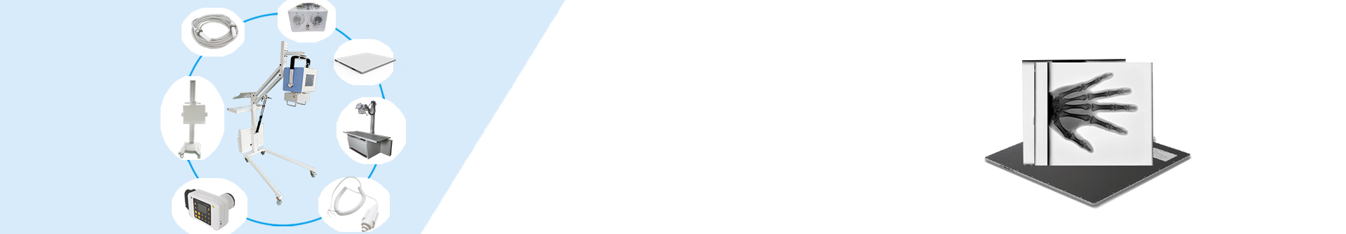
Digital Radiography (DR) flat panel detectors have revolutionized the field of medical imaging. These advanced detectors have greatly enhanced the efficiency and accuracy of medical diagnosis, allowing for clearer and more detailed images of internal body structures. In particular, dynamic DR flat panel detectors have played a crucial role in improving the imaging process, providing real-time visualization of moving anatomical structures. In this article, we will explore how dynamic DR flat panel detectors work and the impact they have had on medical imaging.
Dynamic DR flat panel detectors are a type of digital radiography technology that is designed to capture high-quality, real-time images of moving body parts. Unlike traditional X-ray film or computed radiography (CR) systems, which rely on physical image plates to capture and process images, DR flat panel detectors use a direct digital capture method. This allows for immediate image acquisition and eliminates the need for film processing, resulting in faster imaging times and improved workflow efficiency.
One of the key features of dynamic DR flat panel detectors is their ability to capture images in real-time, making them highly effective for imaging moving anatomical structures such as the heart, lungs, and joints. This is particularly beneficial for procedures such as angiography, fluoroscopy, and orthopedic imaging, where visualization of dynamic processes is essential for accurate diagnosis and treatment planning.
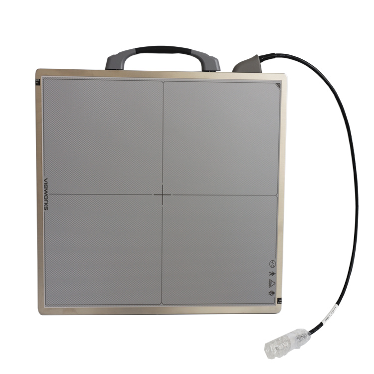
So, how do dynamic DR flat panel detectors work? These detectors consist of a flat panel imaging sensor, which is composed of a scintillator layer and an array of photodiodes. When X-rays pass through the body and strike the sensor, the scintillator layer converts the X-ray energy into visible light, which is then detected and converted into digital signals by the photodiodes. This process allows for the creation of high-resolution digital images that can be viewed in real-time on a computer monitor.
The real-time imaging capabilities of dynamic DR flat panel detectors have had a significant impact on medical imaging practices. By providing immediate visualization of moving anatomical structures, these detectors have improved the accuracy of diagnostic procedures and facilitated more effective treatment planning. For example, in cardiology, dynamic DR flat panel detectors have enabled physicians to visualize the flow of blood through the coronary arteries in real-time, helping to identify blockages and guide interventional procedures with greater precision.
Furthermore, the high sensitivity and dynamic range of dynamic DR flat panel detectors allow for the capture of detailed images with minimal radiation exposure. This is a crucial benefit for both patients and healthcare professionals, as it minimizes the risk of radiation-related complications while ensuring optimal image quality.
In conclusion, dynamic DR flat panel detectors have transformed the field of medical imaging by providing real-time visualization of moving anatomical structures. Their advanced digital capture technology and real-time imaging capabilities have significantly improved the accuracy and efficiency of diagnostic procedures, ultimately leading to better patient outcomes. As technology continues to advance, it is clear that dynamic DR flat panel detectors will continue to play a critical role in shaping the future of medical imaging.
Post time: Aug-26-2025