
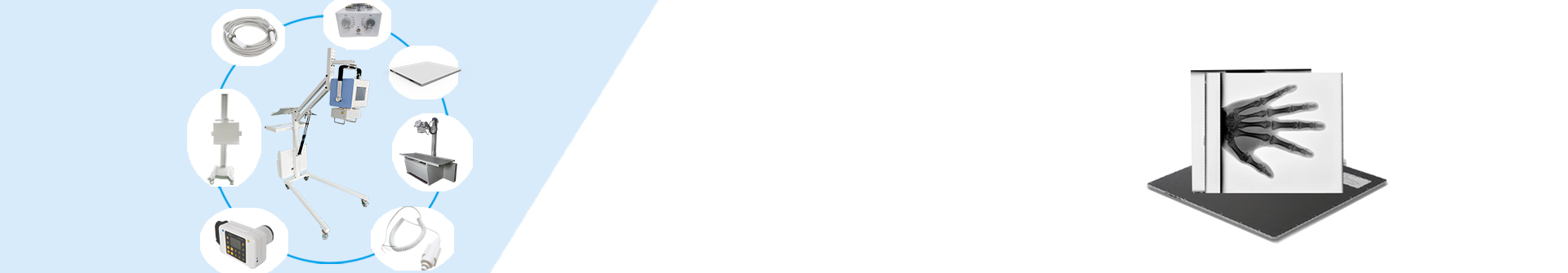
The digital X-ray machine, commonly known as DR, is widely used in medical diagnosis for its convenience. Equipped with a flat-panel detector, it allows direct image viewing on computers, avoiding the trouble of traditional X-ray film development. How does it image? Let’s explore its core principles.
The flat-panel detector is the core of DR, with its material determining imaging performance. There are three main types: Cesium Iodide (CsI), Amorphous Selenium (a-Se), and CCD. Their imaging principles are as follows:
1. Cesium Iodide (CsI) Type
Principle: Indirect conversion. X-rays are first converted to visible light by cesium iodide, then to electrical signals by photosensitive elements, and finally to digital signals via A/D converter for computer imaging.
Feature: High conversion efficiency, good spatial resolution, widely used in general radiography (chest, orthopedics, abdomen).
2. Amorphous Selenium (a-Se) Type
Principle: Direct conversion. Amorphous selenium directly converts X-ray photons to electrical charges, which are read by TFT array and converted to digital signals for imaging.
Feature: High resolution and abundant details, suitable for high-precision diagnosis (mammography, dentistry).
3. CCD Type
Principle: X-rays are converted to visible light by intensifying screen, then collected by CCD sensor to electrical signals, which are digitized for computer processing.
Feature: Mature technology, low cost; slightly lower resolution, gradually replaced in high-end equipment.
Weifang Newheek Electronic Technology Co., Ltd. is a professional X-ray product manufacturer, focusing on DR and flat-panel detector R&D, production and sales, providing high-quality products and technical support.
For more information on technical parameters, application scenarios or quotations, contact us: Tel: +8619953639012. We will provide detailed answers and solutions.
Post time: Dec-19-2025