

At two o’clock in the morning on an orthopedic ward, the patient in bed 3 with multiple injuries suddenly complained of severe pain. The clinician’s first instinct was to order an X‑ray scan to check whether the fractured bone ends had shifted. Yet a tough challenge followed. This patient suffered a femoral fracture and had just undergone skeletal traction. Even slight movement of the leg could displace the fracture ends and damage surrounding soft tissues, undoing the previously satisfactory fracture alignment.
Transferring the patient to the radiology room carried huge risks. Every movement of the patient was a gamble for secondary injury. For fracture patients, the most dangerous journey is often the dozens of meters from the hospital bed to the examination room. What is the solution? Instead of moving the patient, bring the imaging device to them.
How does the wheeled radiography unit work
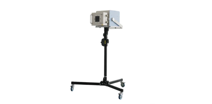
Mobile DR, short for Mobile Digital Radiography System, can be understood as a compact radiology suite that can be wheeled to wards, operating rooms and corridors. Its main components are straightforward: An X‑ray tube generates X‑rays. A flat panel detector converts the rays passing through the human body into digital images. A mobile stand with built‑in batteries supports free movement of the whole unit. A wireless module transmits images to the PACS system in real time.
The full workflow is as follows: The device is wheeled beside the patient’s bed. The tube head and detector are positioned properly. Exposure is performed at bedside. Images are generated within seconds. Clinicians review the images remotely. The patient remains on the bed throughout the entire exam. Traction frames and monitoring lines stay untouched.
Bedside imaging delivers far more than simple radiographs
Many practitioners regard mobile DR merely as an X‑ray machine moved next to beds. In clinical practice, its value extends well beyond that.
Continuous tracking of fracture reduction and healing
It verifies the quality of reduction, changes in bone alignment, stability of implants and callus formation. From emergency reduction to postoperative follow‑up, bedside radiography eliminates risky patient transport just for imaging checks.
Safety verification for traction and fixation hardware
It monitors the position of skeletal traction pins and internal conditions beneath casts or braces. Tiny deviations visible on images can lead to failed reduction outcomes. Bedside imaging enables prompt inspection without removing traction hardware for transfer.
Instant intraoperative and postoperative feedback
During orthopedic reduction or foreign body removal procedures, surgeons require immediate image confirmation. Mobile DR enables seamless cycles of imaging, assessment and continued operation inside surgical suites, without halting surgical workflows.
Radiation safety concerns raised by family members
During bedside scans, family members frequently ask whether nearby exposure leads to excessive radiation. Three key points clarify this concern. First, examinations are only arranged with valid clinical indications and comply strictly with clinical standards. Scans will not be performed unnecessarily. Second, the exposure field is limited strictly to the target area. Lead shielding is applied to radiosensitive organs such as the thyroid and gonads. Third, digital imaging offers superior dose efficiency compared with traditional film‑screen systems. It delivers diagnostic quality at lower patient radiation doses, and scattered radiation can be kept at minimal levels on many standard configurations.
In short, scans are only conducted when clinically necessary, with standardized shielding protocols in place. The core principle of bedside imaging is to bring equipment to the patient, rather than forcing vulnerable patients to travel for scans.
From stationary radiography rooms to every bed inside intensive care units, mobile DR reshapes the fundamental logic of diagnostic imaging in critical care. It lowers transfer risks, speeds up imaging acquisition and helps clinicians make faster, more informed treatment decisions.
 +86 17616362240
 aide@newheekxray.com
How to buy your products ?Do you have distributor in our country?
You can buy the products from our company directly.Normally the procedure is:Sign the contact ,payment byT/T, contact the shipping company to delivery the goods to your country.
What is the warranty?
The free warranty is one year from the day of Commissioning qualified.
Can we visit your country ?
Of course,we will prepare for everything in advance if it is necessary .Generally,it is require that customers must have build agency relationship or business relationship with our company.
How long is the validity of quotation?
Generally,our price is valid within one month from the date of quotation .The price will be adjusted appropriately according to the price fluctuation of raw material and changes in market .
Do you have the CE certificate ?
Yes, most of our products have the CE certificate.
Post time: Nov-16-2026