

Chest radiography ranks among the most routine examinations in radiology departments. Many technologists struggle with a practical question in daily workflows: whether an anti-scatter grid is mandatory for chest exposures. Imaging with and without a grid shows distinct gaps in tonal gradation, lesion detectability and exposure settings. Understanding these differences helps select the proper workflow according to patient physique and equipment conditions.
Origins of Scatter Radiation in Chest Imaging
As X-rays penetrate thoracic structures, tissues of varying density including the thoracic wall, lung parenchyma and mediastinum generate substantial scattered radiation. Scatter photons travel in random directions and create uniform haze over the detector surface, which degrades image contrast. Scatter becomes far more pronounced for patients with larger body mass and thicker chest walls, and this factor serves as the primary criterion to decide grid deployment.
Image Performance Without Anti Scatter Grid
Grid-free acquisition brings less attenuation of primary radiation. Under identical technical settings, more photons reach the detector, allowing lower mAs values, reduced patient radiation exposure and faster capture. For slim individuals with thin chest walls, scatter production stays minimal, and acceptable images can be obtained without a grid. This approach fits paediatric patients and lean adults.
Its drawbacks are equally obvious. For patients with thick chest walls, pervasive haze appears on the image. Lung markings behind the mediastinum and cardiac silhouette turn blurred. Tiny nodules and faint infiltrates may be hidden by fog, raising the risk of missed findings. Overall contrast remains limited, with poor demarcation between bone and soft tissue, making it unsuitable for complicated diagnostic cases.
Image Performance With Anti Scatter Grid
An anti-scatter grid absorbs off-angle scattered photons via lead strips and only transmits primary X-rays aligned with the central beam, effectively eliminating radiographic haze. After grid activation, chest radiograph contrast improves greatly. Lung textures, rib margins and mediastinal contours are well-defined. Lesions in concealed zones such as retrocardiac and subdiaphragmatic regions become easier to identify. This method is ideal for overweight patients, older subjects and cases requiring precise evaluation of pulmonary abnormalities.
The trade-off is partial absorption of useful primary radiation by the grid. Exposure parameters must be increased to maintain adequate brightness, which leads to a moderate rise in patient radiation dose. Additionally, focused grids demand strict control of SID and beam centering. Misalignment creates peripheral cut-off and dark edges, calling for careful positioning by technologists.
Core Comparison of Two Acquisition Strategies
Grid-free imaging: low radiation dose, high positioning tolerance, suitable for patients with thin chest walls. Limited contrast, inadequate image quality for thick body parts.
Grid-assisted imaging: high contrast, rich anatomical details, reliable imaging for thick anatomy and superior diagnostic confidence. Higher radiation output, precise beam alignment required, commonly paired with upright chest stands.
Practical Clinical Guidance
A stratified imaging protocol is recommended instead of universal grid application. Paediatric patients and lean adults should be examined without a grid to minimise radiation burden. Grid usage is advised for overweight adults, pulmonary follow-up scans and cases with suspected nodules or inflammatory changes to guarantee diagnostic clarity.
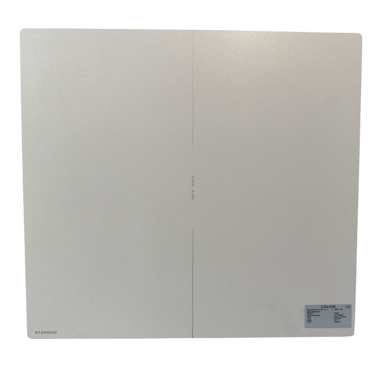
For hardware matching, upright chest stands normally adopt focused grids with 180 cm focal distance and 10:1 grid ratio, a mature configuration for chest radiography. For bedside DR chest imaging where SID cannot be fixed, parallel grids are preferred to avoid cut-off artefacts caused by focal distance mismatch.
The core principle of chest radiography is not a rigid rule to use or skip the grid. Patient chest thickness should act as the main reference, balancing diagnostic image quality and radiation protection. Proper workflow selection avoids unnecessary radiation while ensuring images meet diagnostic requirements.
Whatsapp:+86 17616362240
aide@newheekxray.com
Post time: Nov-10-2026