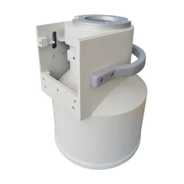
Image intensifiers are vital tools in medical imaging, amplifying low-intensity X-rays into bright, clear visuals for real-time diagnostics. Commonly used in fluoroscopy, these devices enable surgeons to monitor procedures like angiography or orthopedic interventions with precision.
The technology works by converting X-ray photons into electrons via a photocathode, accelerating them through an electric field, and projecting the amplified image onto a fluorescent screen. This process enhances brightness up to 10,000 times, allowing lower radiation doses for patients while maintaining image quality.
Modern advancements include digital integration, where intensifiers pair with CCD/CMOS sensors to produce high-resolution digital videos, streamlining workflows in emergency and surgical settings. Compact designs also support portable C-arm systems, ideal for mobile clinics or operating rooms.
Though gradually supplemented by flat-panel detectors, image intensifiers remain cost-effective solutions for dynamic imaging, particularly in resource-limited settings. Their blend of reliability, affordability, and real-time capability continues to make them indispensable in modern healthcare.
Post time: May-08-2025


