
The image intensifier was born in the 1950s and was a great product. His appearance ended the history of screen imaging. It made the X-ray fluoroscopy dose greatly reduced in that era, the convenience of the technician was greatly improved, and the patient and the technician received a greater degree of protection.
Similarly, with the development of technology, image intensifiers have come to today, and they have gradually entered old age, and the fate of being replaced has long been arranged. With the breakthrough of various dynamic image technologies, image intensifier imaging technology is gradually eliminated.
Today, I will not cherish the memory of the image intensifier here, but only analyze why the image intensifier was eliminated with everyone. I think there are mainly a few reasons:
First: the imaging format is small, and it is easy to miss and misdiagnose.
As can be seen from the figure below, the left side is an image formed by an imaging augmentation of the whole digestive tract, which can only contain part of the inspected part in one frame; the right side is the current mainstream large-scale imaging, which can contain the whole The entire inspection site of the digestive tract can be more convenient for observation and diagnosis.
Under normal circumstances, when using contrast enhancement imaging, it is necessary to continuously move the position of the shadow enhancement, follow the flow direction of the contrast agent, and conduct real-time observation, so as to better capture the lesion point, but for the inspection with a faster contrast agent flow rate, it is easy to The device cannot keep up with the movement, so it cannot be observed. For example, in esophagography, it is easy to appear the phenomenon of contrast increase and dislocation of contrast agent.
The small imaging format has become a very important reason for the limited development of image augmentation. So, is it possible to make the shadow bigger? In fact, it can be seen from the working principle of the shadow increase that with the increase of the imaging format, the volume of the entire shadow increase also changes greatly, and eventually it cannot be used in coordination with the whole machine, so the current largest shadow increase can only reach 12 inches, commonly used The main ones are 7/9inch.
Second, it is easy to be distorted and distorted, and it is easy to be missed and misdiagnosed.
Due to its working principle, image intensifiers are prone to distortion and distortion. Distortion There are two main types of distortion: one is circular balanced geometric distortion; the other is asymmetrical, commonly referred to as S-distortion.
The reason for the geometric distortion is that the projection of the X-ray image onto a curved surface produces a larger image of an object on the entrance plane at the edges of the input screen than in the middle. This distortion is related to the geometry of the input screen and the variation of the X-ray source. position-dependent, so it is called geometrical distortion. A lens with negative distortion will partially compensate for the positive distortion due to the curvature of the input screen, thus reducing the overall distortion of the output image, but the distortion cannot be avoided.
Another type of distortion is called S-distortion, which is due to the characteristic S-shaped image of rectilinear objects, a phenomenon caused by interference from the Earth’s magnetic field or stray magnetic fields from surrounding equipment.
It is precisely because of the distortion and distortion (as shown in the figure below) that it seriously interferes with the diagnostic inspection results of X-ray images, which may easily lead to missed diagnosis and misdiagnosis.
Third, the image contrast is low, which is easy to be missed and misdiagnosed.
At present, the dynamic range of mainstream X-ray imaging is 14-bit or 16-bit, while the dynamic range of image intensifier is only 10-bit. In other words, the dynamic range of the current mainstream dynamic imaging products is 16 times or 32 times that of the film.
The dynamic range is different, and the result is as shown in the figure below. The dynamic range on the left is obviously much worse than that on the right, so the fineness and color of the image are very different.
The image of the shadow increase is shown in the figure below. The dynamic range of 10 bits will be helpless in the observation of lesions with small differences in image density, especially in the exudative and diffuse imaging pathological changes such as early SARS lung changes. It cannot be diagnosed correctly, which can easily lead to missed diagnosis and misdiagnosis.
Technology is changing with each passing day, and product changes are earth-shaking. Image intensifiers have gone through their glorious days and have reached the end of their lives. There are bound to be more breakthroughs in medical imaging diagnosis. Remembering the past and looking forward to the future, everything will eventually become history.
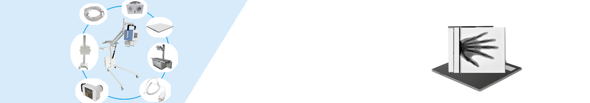
If you are interested in our products, welcome to consult.
Post time: Aug-20-2025