
As the “X-ray eye” for oral diagnosis and treatment, dental X-ray machines use low-dose X-rays to help dentists detect problems that cannot be detected by the naked eye, ensuring accurate diagnosis and treatment. Modern technology has significantly reduced radiation risks, and regular dental checkups are key steps in maintaining oral health. It is generally recommended that adults undergo a check-up every 1-2 years, and children and high-risk individuals need to pay more attention to scientific diagnosis in order to prevent problems before they occur.
Fundamentals and Types of Dental Slices Machines
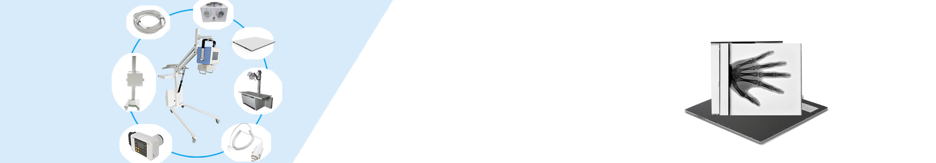
Dental X-ray machine, also known as dental X-ray machine, is an indispensable imaging diagnostic equipment in oral diagnosis and treatment. It emits low-dose X-rays to “see through” the internal structures of teeth and jawbones, helping dentists discover hidden problems such as dental caries and root apex lesions, and compensating for the shortcomings of visual observation. This device plays a crucial role in routine inspections, allowing problems to be exposed early and preventing small issues from escalating into serious illnesses. Dentists use it for comprehensive evaluation to ensure the scientific and personalized treatment plan, thereby improving overall oral health. There are various types of dental film machines to meet different diagnostic needs, among which intraoral dental film machines are the most commonly used, used to capture single or several teeth, especially suitable for examining local problems such as dental caries and periodontal conditions. The panoramic camera (OPG) can capture images of all teeth and upper and lower jaw bones, which can be used in wisdom tooth assessment or implant planning. Cone beam CT (CBCT), as a three-dimensional imaging device, provides a three-dimensional view of teeth, neural tubes, etc., and is widely used in complex preoperative planning. These types collectively cover the diverse scenarios of oral diagnosis and treatment.
Working principle and safety analysis
The working principle of dental film machine is based on the difference between X-ray penetration and absorption. The device first generates a beam of radiation through an X-ray tube. When passing through oral tissue, tissues of different densities such as teeth and bones will affect the radiation path with varying degrees of absorption. High density parts such as dentin absorb more radiation, forming bright areas; However, soft tissues absorb less and appear as dark areas. This process captures the signal after penetration through a detector, converts it into preliminary image data, and then finely processes and displays a clear view through computer algorithms, helping dentists intuitively identify internal structural abnormalities. The radiation dose of modern dental examination is extremely low, and its safety is fully guaranteed. For example, the radiation dose of a small dental plaque is only about 0.005mSv, which is equivalent to taking a 2-hour flight or being exposed to natural environmental radiation for a day; The panoramic film is only 0.01-0.02mSv, far below the harmful threshold of 100mSv/year. Equipment design and protective measures further reduce risks, and dentists strictly control exposure time and range during operation. This low-dose characteristic ensures the safety of frequent examinations, encourages people to receive regular diagnosis, and prevents the deterioration of oral diseases.
Diagnostic ability and discovery
Dental film machine is a powerful tool for discovering hidden problems, especially adept at exposing dental caries that cannot be recognized by the naked eye, such as secondary caries between teeth or under existing fillings, which are often overlooked due to their hidden location. The device can also clearly display apical lesions, including apical abscesses or granulomas, and provide early warning through image evaluation of bone changes. In addition, the diagnosis of periodontal disease benefits from the visualization of the degree of alveolar bone resorption, which helps dentists assess inflammation progression and bone health, avoiding tooth loosening caused by periodontal deterioration. Dental film machine plays a significant role in the diagnosis of complex problems, accurately locating the growth direction and position of obstructed teeth, such as abnormal emergence of wisdom teeth, reducing surgical risks. It can also reveal jaw lesions such as cysts or tumors, and guide early intervention through image features. Abnormal tooth development cannot escape its “eyes”, such as problems with supernumerary or missing teeth, which can be corrected in a timely manner to avoid functional defects. This comprehensive diagnostic capability enables dentists to develop personalized treatment plans, supported by reliable evidence from orthodontics to surgical procedures.
Advantages of digital technology
The digital dental film machine brings revolutionary progress in radiation control, reducing the dose by 50-80% compared to traditional film, further improving safety. This technology also enables fast imaging, displaying high-resolution images within seconds and reducing patient waiting time. Images can be presented in real-time through computer screens and support post-processing, such as adjusting contrast or zooming in on details to enhance diagnostic accuracy. Its convenience is reflected in the real-time playback function, allowing dentists to confirm the results on the spot and communicate. Storage and transmission efficiency is another major advantage of digital dental films. Images are saved in electronic format, which facilitates long-term management and retrieval in databases, avoiding damage or loss of physical films. It also supports remote transmission to experts or cloud platforms, promoting collaborative diagnosis and treatment. In terms of environmental protection, there is no need for chemical washing process, reducing harmful waste emissions, which is in line with the concept of green medicine. These improvements significantly reduce clinic operating costs, enable advanced technology to benefit more patients, and enhance the overall medical experience.
Precautions for Photography and Special Groups
When taking dental photos, attention should be paid to details to ensure quality and safety, such as removing metal objects such as jewelry to prevent interference with the image. The patient needs to cooperate with the technician to adjust their position and maintain absolute stillness, as any movement may cause image blurring; Special groups such as children can use protective equipment or specialized low-dose modes. It is recommended to avoid taking repeated shots of the same area in the short term to control accumulated radiation. These measures not only improve image clarity, but also ensure individual safety. For pregnant women, dental examination should be suspended in non emergency situations; When inspection is necessary, a lead apron should be used for tight protection. Thyroid disease patients may request to wear thyroid protection collars to reduce exposure. Children and the elderly are sensitive groups, and the equipment is equipped with exclusive settings to optimize safety. It is crucial to inform the doctor of the medical history in advance to avoid risks. These targeted strategies embody medical humanistic care, ensuring reliable diagnosis under different needs and encouraging the public to accept necessary examinations with confidence.
Future Trends and Health Suggestions
Dental film machines are developing towards lower doses and intelligence, with increased sensor sensitivity to further compress radiation and protect patient safety. Artificial intelligence assisted diagnosis is gradually becoming popular, which can automatically identify lesions such as dental caries or bone resorption, improving efficiency. The miniaturization and cost reduction of 3D imaging devices such as CBCT have made them accessible to routine clinics. Chair based real-time imaging technology integrates treatment equipment to shorten diagnosis and treatment time. These trends make high-tech diagnostics more accessible and widespread. To maintain oral health, it is recommended to develop a regular plan in conjunction with dental checkups: adults should have their teeth checked every 1-2 years, and children and high-risk individuals such as those with high tooth decay rates should have their teeth checked more frequently. Pay attention to daily oral hygiene, but brushing teeth alone is not enough to detect deep-seated problems in a timely manner. Following the doctor’s advice to take dental X-rays is crucial to avoid delaying treatment. Future remote diagnosis and treatment support real-time image sharing, facilitating expert consultations. Scientific examination is the key to prevention, ensuring a healthy and long-lasting smile.
Post time: Jun-09-2025