
Why do we need dental X-rays?
Many dental problems are like an iceberg – only 10% of the surface is exposed, and 90% is hidden under the gums! Our dental X-ray machine has discovered:
✔️ Hidden dental caries (especially adjacent caries)
✔️ Root inflammation and cyst
✔️ Direction and position of wisdom tooth growth
✔️ Alveolar bone resorption status
✔️ Preoperative evaluation of dental implantation
Three common dental X-ray examination methods
1.Root tip slice
Checking the details of a single tooth like a “close-up shot of teeth” takes about 30 seconds
2. Panoramic film (curved fault)
One shot full mouth dental image “Family portrait of teeth” suitable for orthodontic and implant planning
3.CBCT 3D imaging
Obtain three-dimensional dental imaging images and accurately measure bone mass through “3D modeling of teeth”
The 3 most concerning issues for patients
Q: Is the radiation level high?
A: A dental X-ray examination radiation ≈ the natural radiation dose of eating 10 bananas Q: Can pregnant women take photos?
A: If necessary, protective clothing can be worn. Please consult your dentist Q: How often is appropriate for taking photos?
A: Routine examinations should be conducted once every 1-2 years, and treatment should be followed as needed
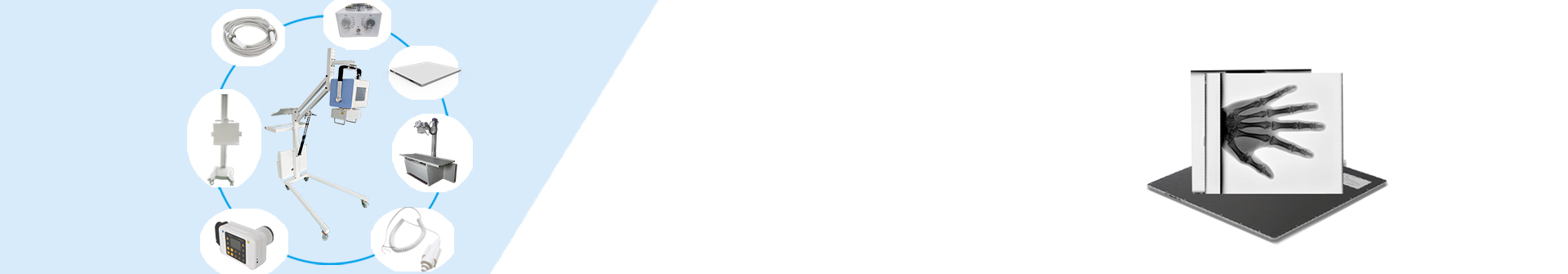
X-ray system
✅ 0.05 second high-speed imaging – even pediatric patients can easily cooperate
✅ AI Image Optimization System – Automatically Identify 20+Common Dental Diseases
✅ Nano level protection design – radiation level is only one-third of the international standard
✅ 5G remote consultation module – real-time sharing of high-definition images
Post time: Jun-24-2025