

When it comes to diagnosing problems related to the chest area, medical professionals often rely on two imaging techniques: chest X-ray and chest CT. These imaging modalities play a crucial role in detecting various respiratory and cardiac conditions. While both are essential tools, it is essential to understand the differences between them to ensure accurate diagnoses and effective treatments.
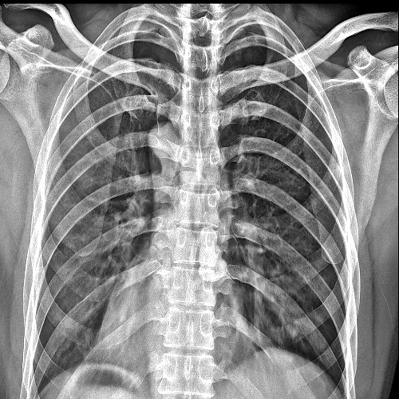
A chest X-ray, also known as a radiograph, is a commonly used imaging technique that produces a static image of the chest using electromagnetic radiation. It involves exposing the chest area to a small amount of ionizing radiation to capture images of the lungs, heart, blood vessels, bones, and other structures. Chest X-rays are cost-effective, readily available, and provide a quick overview of the chest region.
On the other hand, a chest CT scan, or computed tomography, utilizes a combination of X-rays and computer technology to produce cross-sectional images of the chest. By generating multiple detailed images from different angles, a CT scan provides an in-depth view of the chest, highlighting even the tiniest abnormalities. CT scans are particularly useful in diagnosing complex conditions and analyzing the chest’s internal structures.
One significant difference between a chest X-ray and a chest CT lies in their imaging capabilities. While both techniques allow for the visualization of organs and tissues within the chest, a chest CT provides a much higher level of detail. A chest X-ray offers a broad overview but may not reveal smaller abnormalities or subtle changes in tissues. On the contrary, a chest CT can detect and characterize even the most intricate structures, making it more useful in identifying specific conditions.
The clarity and precision of a chest CT scan make it an invaluable tool in diagnosing various respiratory and cardiac conditions. It can identify lung cancer, pulmonary embolism, pneumonia, and evaluate the extent of lung damage caused by diseases such as COVID-19. Additionally, chest CT scans are often used in individuals with suspected heart conditions, providing detailed images of the heart and surrounding blood vessels to detect abnormalities, such as coronary artery disease or aortic aneurysms.
While a chest CT scan offers exceptional imaging capabilities, it is not always the initial imaging choice. Chest X-rays are typically performed as the first-step screening tool due to their affordability and accessibility. They are often used to identify common chest abnormalities and guide further diagnostic investigations, such as CT scans or other imaging modalities.
Another key difference between a chest X-ray and a chest CT is the level of radiation exposure. A typical chest X-ray involves minimal radiation exposure, making it relatively safe for routine use. However, a chest CT scan exposes the patient to a higher dose of radiation due to the multiple X-ray images taken throughout the procedure. The risk associated with radiation should be carefully weighed against the potential benefits of a chest CT scan, particularly in pediatric patients or individuals requiring multiple scans.
chest X-rays and chest CT scans are vital diagnostic tools used in the evaluation of respiratory and cardiac diseases. While a chest X-ray provides a basic overview of the chest area, a chest CT scan offers detailed and precise images, making it ideal for identifying complex conditions. The choice between the two depends on the specific clinical context, availability, and the level of detail required for an accurate diagnosis.
Post time: Oct-30-2023