
I. Basic Properties of X-Rays
X-rays, visible light, and ultraviolet light all belong to the electromagnetic spectrum, but they differ significantly in wavelength and frequency. X-rays have an extremely short wavelength (shorter than the diameter of an atom), which gives them strong penetrating power. At the same time, due to their high energy, they can interact with atoms of matter and ionize them. The charged particles generated by ionization will further react with biomolecules (such as DNA), possibly causing DNA strand breaks or base mutations. This is the core source of the X-ray radiation risk that the public generally concerns about.
II. X-Ray-Based Medical Imaging Technologies
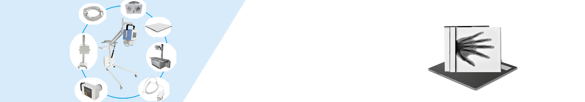
1. Digital Radiography (DR)
DR (Digital Radiography) is a commonly used X-ray imaging device in clinical practice. Its core components and functions are as follows:
X-ray tube: The core emitting component that generates X-rays under the action of high voltage.
X-ray high-voltage generator: Provides a stable high-voltage electric field for the X-ray tube to ensure the continuous and stable generation of X-rays.
Flat-panel detector: A signal-receiving component that replaces traditional film, which can convert X-ray signals passing through the human body into digital electrical signals.
Mechanical components: Including racks, examination beds, etc., used to adjust the scanning angle and the patient’s position to ensure the accuracy of the imaging position.
Imaging system: Processes and reconstructs the digital signals transmitted by the flat-panel detector, and finally generates clear digital images.
The imaging principle of DR is based on the “difference in X-ray absorption by human tissues”. When X-rays pass through the human body, high-density tissues (such as bones) have a strong ability to absorb X-rays, resulting in a small amount of penetrating rays and weak signals received by the flat-panel detector, which appear as bright areas in the image. In contrast, soft tissues (such as muscles and internal organs) have weak X-ray absorption, allowing more penetrating rays, and thus appear as darker areas in the image. This “planar imaging” mode is equivalent to compressing the human structure onto a two-dimensional plane, reflecting the difference in tissue density through the contrast of light and dark.
The core advantages of DR lie in its relatively low cost and simple operation, making it suitable for quickly completing routine examinations. However, it should be noted that it still poses a risk of ionizing radiation, and strict compliance with radiation protection standards is necessary.
2. Computed Tomography (CT)
CT (Computed Tomography) is a three-dimensional imaging technology developed on the basis of traditional X-ray imaging. Its emergence stems from the need to overcome the limitations of two-dimensional imaging. Traditional X-ray films (and DR) can only present planar images, which cannot clearly show the three-dimensional structure of tissues. CT achieves three-dimensional imaging through the following methods:
Scanning phase: The X-ray emitting device and detector rotate around the human body, acquiring a large number of two-dimensional tomographic images (each layer can be as thin as 0.5mm) from multiple angles (usually rotating dozens of times per second).
Reconstruction phase: A computer algorithm is used to superimpose and calculate all two-dimensional tomographic images to reconstruct a three-dimensional image of human tissues, which can clearly show the structural details of different layers.
Similar to DR, the brightness of CT images is also determined by tissue density. High-density tissues such as bones appear bright in CT images, while fat and soft tissues appear as medium to low brightness. Especially in foreign body localization, since foreign bodies (such as metal fragments and ingested objects) usually have much higher density than human tissues, they can be clearly visualized in CT images, facilitating accurate localization.
III. Clinical Application Advantages of X-Ray Imaging
X-ray imaging technologies (DR, CT) have strong resolution capabilities for high-density substances, so they are mainly used in clinical examinations for the following purposes:
Skeletal system examination: Clearly display the location, scope, and severity of lesions such as fractures, bone tumors, and osteoporosis.
Spine and joint examination: Observe organic lesions such as scoliosis, intervertebral disc herniation, and joint degeneration, and clarify the relationship between the lesions and surrounding soft tissues (such as nerves and muscles).
Foreign body localization: Quickly locate foreign bodies in the body (such as metal fragments and ingested foreign bodies) to provide an accurate basis for surgical removal.
Partial chest and abdominal examinations: Assist in the diagnosis of lesions such as pneumonia, pulmonary tuberculosis (DR), pulmonary nodules (CT), and abdominal effusion, providing an imaging basis for subsequent diagnosis and treatment.
Post time: Sep-02-2025