
First, we clarify the key differences between high-frequency X-ray machines and power-frequency X-ray machines, then compare the operational efficiency of DR (Digital Radiography) with traditional X-ray machines and CR (Computed Radiography), and finally elaborate on DR’s unique advantages.
1. Core Advantages of High-Frequency vs. Power-Frequency X-Ray Machines
High-frequency X-ray machines outperform power-frequency models in multiple critical aspects, making them more suitable for modern clinical needs:
- Stable output & high imaging quality: High-frequency machines adopt high-frequency inversion technology, enabling stable X-ray output with low ripple (usually <5%). This reduces image noise and ensures clear, detailed imaging. In contrast, power-frequency machines have high ripple (20-40%), leading to greater image distortion.
- Low radiation dose: High-frequency machines achieve efficient X-ray conversion (energy conversion rate >80%), so they require lower tube current/voltage to obtain qualified images—reducing radiation exposure to patients and medical staff by 30-50% compared to power-frequency machines.
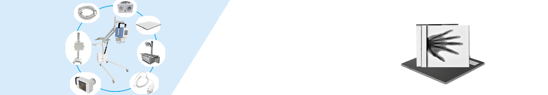
- Compact size & flexible installation: High-frequency generators are smaller and lighter (1/3 the size of power-frequency generators), making them ideal for mobile bedside machines or limited-space radiology rooms. Power-frequency machines are bulky and require fixed installation due to their heavy transformers.
- Fast response & energy savings: High-frequency machines have a shorter exposure preparation time (≤0.5s) and lower energy consumption (saving ~30% electricity vs. power-frequency models), improving operational efficiency and reducing hospital costs.
2. Operational Efficiency Comparison: DR vs. Traditional X-Ray Machines & CR
In terms of time consumption (a key factor in clinical workflow), DR significantly outperforms traditional power-frequency X-ray machines and CR:
- Traditional power-frequency X-ray machines: It takes ~6 minutes to complete the entire process (exposure → film development → film retrieval).
- CR systems: The process (exposure → image plate scanning → image processing) takes ~7 minutes, as it requires additional steps for plate handling and scanning.
- DR systems: Imaging is completed in one exposure (exposure → real-time digital image acquisition). The entire process takes only seconds, drastically accelerating operational efficiency and reducing patient waiting time.
3. Additional Comparisons: DR vs. Traditional X-Ray/CR
Difference 2: Compatibility
DR has excellent compatibility with most clinical scenarios. It works seamlessly with flat-panel detectors of standard sizes and can adapt to basic imaging needs (e.g., chest, limbs, spine) in primary hospitals, clinics, and tertiary hospitals—making it a versatile choice for universal use. Traditional X-ray machines and CR, by contrast, have limited compatibility (e.g., CR relies on specific image plate sizes, and old power-frequency machines may not support digital upgrades).
Difference 3: Operability
DR features user-friendly design: Most models are equipped with touchscreens, pre-set anatomical programs, and one-click exposure functions. This simplifies operation, reduces the learning curve for staff, and saves time/labor—directly improving daily work efficiency. Traditional X-ray machines require manual adjustment of multiple mechanical knobs, while CR adds steps like plate calibration, increasing operational complexity.
4. Key Advantages of DR (Digital Radiography)
DR integrates advanced digital technology, bringing revolutionary improvements to imaging workflows and diagnostic capabilities:
- Wide dynamic range & exposure latitude: DR tolerates minor technical deviations during imaging (e.g., slight errors in exposure parameters). Even for body parts where exposure control is difficult (e.g., chest with overlapping tissues), it still produces high-quality images—reducing re-examination rates.
- High resolution & rich image information: With high spatial resolution (up to 3.5 LP/mm) and a wide grayscale range (16-bit, supporting 65,536 gray levels), DR captures fine anatomical details (e.g., small lung nodules, subtle bone fractures). Paired with advanced digital image algorithms, it fully guarantees image quality for accurate diagnosis.
- Real-time fluoroscopy & flexible post-processing: In fluoroscopy mode, DR displays digital images in real time. Doctors can perform targeted photography based on patient conditions, then use post-processing functions (edge enhancement, zooming, black-white inversion, image smoothing) to extract rich clinical information. This is particularly critical for detecting early-stage lesions (e.g., early lung cancer, mild osteoarthritis).
- Low radiation dose: DR’s efficient X-ray conversion and digital image enhancement technology reduce the required radiation dose by 40-60% compared to traditional power-frequency X-ray machines—while still delivering clearer images. This minimizes radiation risks for both patients (especially children and pregnant women) and medical staff.
- Filmless operation & cost savings: DR eliminates the need for film (achieving “filmless radiography”). Patient images are stored digitally in computers, reducing hospital costs for film procurement, storage, and management. It also speeds up image retrieval (no need to search for physical films), improving workflow efficiency.
- PACS integration & telemedicine support: DR seamlessly integrates with hospital PACS (Picture Archiving and Communication Systems), enabling remote expert consultations, online case discussions, and cross-department image sharing. Additionally, Tiandi Smart DR (a featured model) supports multi-image display and side-by-side image comparison—helping doctors identify subtle changes (e.g., before/after treatment) and make accurate diagnoses.
If you are seeking high-quality X-ray machines (including high-frequency models and DR systems) or need further product details, please feel free to contact us.
Post time: Sep-11-2025